BETTER MECHANICS WON'T SAVE YOUR UCL: HERE'S WHY
The short version
"Fix his mechanics" is baseball's most common advice, and a logical fallacy. The research says tissue capacity and workload are what actually matter.
By Dylan Newcomer, PT, DPT
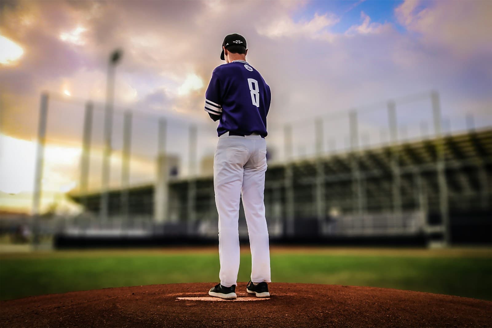
Table of Contents
A 16-year-old throws 101 mph on video. It makes the rounds on X. And within hours, the most predictable thing in baseball happens: adults who do not throw baseballs for a living start diagnosing his mechanics from a cell phone video.
"That arm action is going to get him hurt."
"He needs to clean up his mechanics before he blows out his elbow."
An orthopedic surgeon (someone who should know better) weighs in with the same take. Fix the mechanics, save the UCL.
This is the most common logical fallacy in baseball. And it is worth unpacking, because it is not just wrong. It is the kind of wrong that leads to bad advice being handed to talented kids. Meanwhile, Solomito et al. (2024) analyzed 19,348 UCL injuries in males aged 10 to 34 and found that 54% occurred in the 15-to-19 age group, and UCL injury rates remain elevated despite decades of pitch count rules and mechanics coaching. The current approach is not solving the problem.
The "Better Mechanics" Paradox
Here is what people mean when they say "better mechanics": optimized hip-shoulder separation, efficient trunk rotation, and maximized distal segment velocity. In other words, a more efficient kinetic chain that transfers force from the ground through the trunk and out through the arm.
Here is the problem. That mechanical efficiency does not reduce elbow stress. It increases ball velocity. And ball velocity is a strong correlate of valgus torque at the elbow. The exact force that loads the UCL.
A 2023 qualitative systematic review in Arthroscopy examined 2,896 pitchers across high school, collegiate, and professional levels. Manzi et al. found that pitch velocity was significantly correlated with elbow varus torque, with the strongest association appearing in high school pitchers (R² = 0.36) and collegiate pitchers (R² = 0.29). The professional-level correlation was weaker (R² = 0.076), but the direction was the same, and injured professionals had significantly faster pitch velocity before injury compared with uninjured controls.
The data from the major leagues tells the same story even more bluntly. Mastroianni et al. (2025) compared 115 MLB pitchers who underwent UCL surgery against 230 matched controls and found that higher velocity across all pitch types was associated with surgery. But here is the part that should make everyone uncomfortable: superior Pitching+ scores (a composite metric of overall pitching ability) also correlated with UCL surgery. The best pitchers in the world are the ones most likely to blow out their elbows. A companion analysis by the same group put a number on it: each 1 mph increase in velocity raised UCL surgery risk by approximately 20%. And there were no significant differences in pitch counts, spin rates, release points, or pitch movement between the injured and uninjured groups. The variable that separated them was how hard they threw.
Sakurai et al. (2024) confirmed this at the collegiate level with an important nuance: the relationship is pitcher-specific. Of 81 Division I pitchers, 39 showed significant torque-velocity correlations. The other 42 did not. But even in this study, the within-pitcher relationship (R² = 0.29) explained substantially more variance than the across-pitcher relationship (R² = 0.05), meaning that when a given pitcher throws harder, he tends to generate more torque at the elbow.
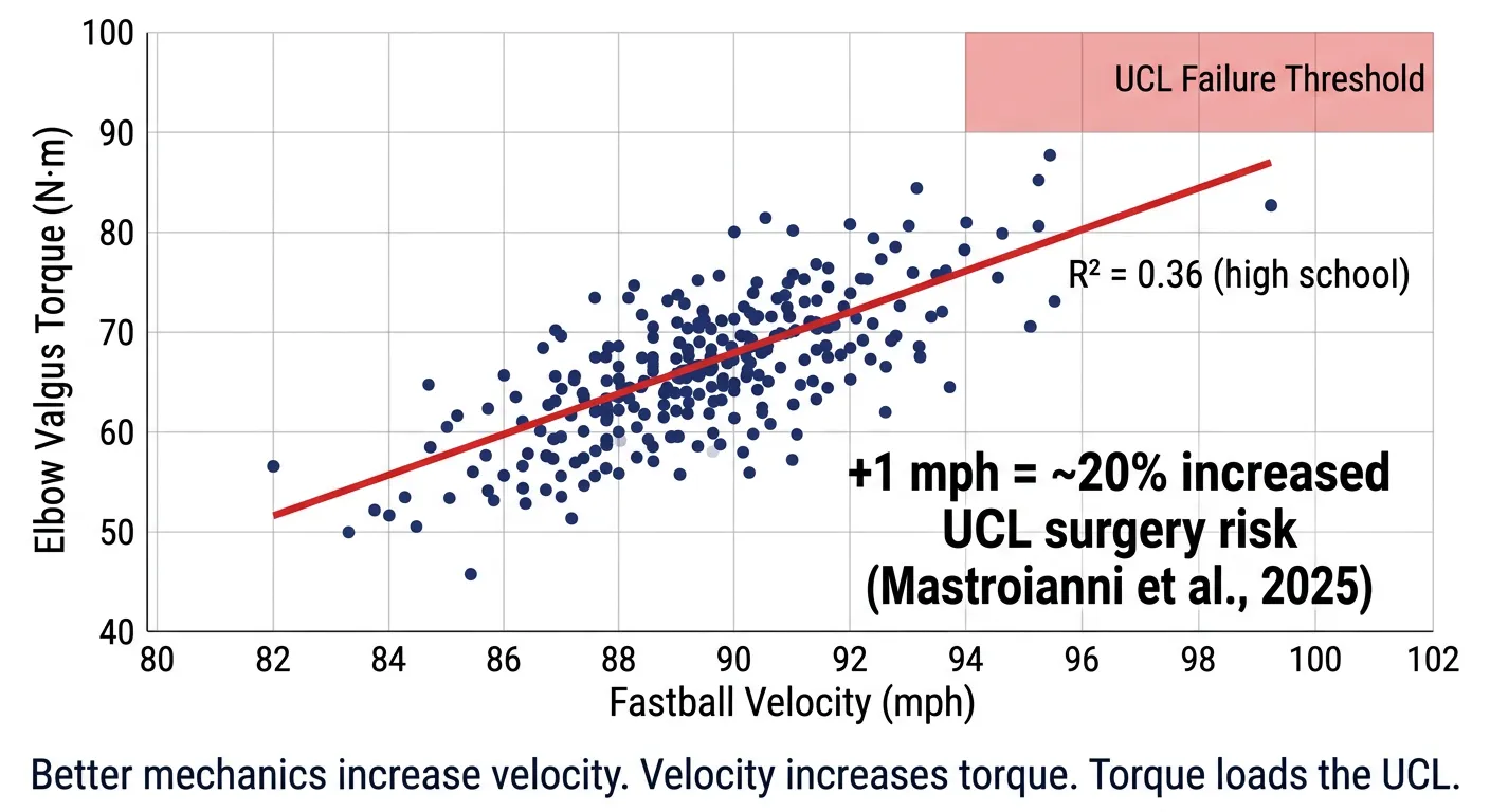 Velocity-torque scatter plot showing a clear positive correlation between fastball velocity and elbow valgus torque, with a UCL failure threshold in the upper right. Each 1 mph increase raises UCL surgery risk by approximately 20%.
What does this mean in plain English? Improving mechanical efficiency without also increasing velocity (and thus likely increasing torque at the UCL) is the exception, not the rule. You cannot optimize the engine and expect the drivetrain to experience less stress. The physics do not work that way.
A kid throwing 101 at 16 is not doing that with bad mechanics. He is already operating in the top 0.01% of mechanical efficiency on the planet. Telling him to "fix his mechanics" is telling him to change something that is already working at an elite level, based on nothing more than someone's subjective preference for how an arm action should look.
Nobody Has Defined "Good Mechanics"
This brings us to the second problem: nobody has ever agreed on or clearly defined what "good mechanics" actually are.
Chalmers et al. (2017) reviewed the relationship between pitching mechanics and injury in Sports Health and arrived at a striking conclusion: "Currently, no prospective clinical evidence exists to connect shoulder proximal force with rotator cuff tears, shoulder rotational forces with proximal humeral epiphysiolysis, or shoulder rotational torques with internal impingement." The evidence linking mechanics to injury is correlational, not causative. They also noted that in a one-year prospective study of 476 youth pitchers with video motion analysis (Lyman et al., 2002), no motion analysis factor could be linked to injury. Not one.
Bullock et al. (2021) reinforced this in a systematic review in the Journal of Science and Medicine in Sport: "Due to the low power of many of these studies, and the lack of prospective 3D biomechanical studies," most biomechanical variables cannot be confirmed as injury risk factors.
So when someone looks at a video of a 16-year-old throwing 101 and says his arm action is going to get him hurt, what they are actually doing is stating a preference. They have decided they do not like the way his arm works. That is not a clinical finding. That is an aesthetic judgment. And aesthetics and mechanical efficiency are not the same thing. Conflating the two is exactly how bad advice gets handed to talented kids.
What Actually Determines Injury
The injury equation is simpler than people want it to be:
Stress Applied > Tissue Capacity = Injury
That is the whole thing. When the cumulative stress placed on a tissue exceeds that tissue's ability to absorb it, the tissue fails. Not because of how the stress was delivered. Because the stress exceeded the capacity.
Fleisig et al. (2025) published the most rigorous prospective study to date, 305 professional baseball pitchers followed over 4.5 years with baseline biomechanical testing. The only significant predictor of UCL surgery was elbow varus torque, with a 26% increase in risk for every 10 N·m increase. Fastball velocity, body size, prior surgery, range of motion deficits, and even UCL abnormalities on baseline MRI were all non-significant. The stress on the tissue was the variable that mattered.
Now, can specific mechanical faults create abnormal stress concentration at specific tissues? Yes, and that is worth acknowledging. Early trunk rotation, excessive elbow drop, or a broken kinetic chain can create localized shear and impingement at specific anatomical locations. Bullock et al. (2021) found that early trunk rotation predicted elevated surgical risk with a hazard ratio of 1.69. Fix the fault, remove that concentrated stressor, and symptoms resolve. That mechanism is real and clinically meaningful.
But it is a localized tissue irritation problem, not a global valgus torque problem. Those are two completely different injury mechanisms, and conflating them is where the argument breaks down. Valgus torque at the UCL scales with velocity. It does not care how pretty the arm action looks.
The Flexor-Pronator Group: Your UCL's Real Protection
If mechanics are not the primary lever for UCL protection, what is?
The answer is the musculature that sits directly on top of the ligament. The flexor digitorum superficialis (FDS), flexor carpi ulnaris (FCU), flexor digitorum profundus (FDP), and pronator teres are the dynamic stabilizers sharing load with the UCL during every throw.
Park and Ahmad (2004) demonstrated in a cadaveric study that combined contraction of the FCU and FDS provided the greatest correction of valgus angle at both 30 and 90 degrees of elbow flexion. Their model identified the FCU as the primary stabilizer and the FDS as secondary. But cadaveric testing isolates muscles in ways that do not capture the full picture of how these structures interact during a live throw, and more recent work has shifted the conversation toward the FDS as the structure that may matter most.
Here is why. Patel et al. (2020) reviewed the anatomy of the medial UCL complex and found that the FDS tendon overlaps 46% of the UCL's ulnar footprint. The FCU overlaps just 21%. The FDS has more than double the anatomical coverage of the ligament it is protecting. Matsuzawa et al. (2021) described the FDS as "the most important of the forearm flexors" for reducing valgus stress and noted that the second and fifth digit FDS tendons actually originate from the anterior bundle of the UCL itself. The muscle is not just near the ligament. Parts of it are physically attached to it.
Hoshika et al. (2020) showed that FDS contraction (specifically of the index and middle fingers) significantly reduced medial joint distance under valgus stress, confirming the dynamic stabilizing role through a tendinous septum mechanism.
And when UCL injuries occur, the FDS is almost always involved. Ikezu et al. (2022) examined 99 baseball players with medial elbow injuries via MRI and found that 45 had combined UCL and flexor-pronator injuries. Of those 45, 40 (89%) involved the FDS specifically, with injuries occurring in the deep layer of the muscle belly. Distal UCL tears were significantly associated with concomitant FDS damage.
Otoshi et al. (2022) demonstrated the clinical consequence from the other direction: in patients with confirmed UCL insufficiency, isometric forearm pronation reduced valgus stress-induced joint widening from 5.1 mm to 2.6 mm and completely eliminated medial elbow pain. The muscles did what the damaged ligament could not.
The literature does not fully agree on which muscle is the single most important stabilizer. Park and Ahmad's cadaveric data points to the FCU. Saito's prospective injury data (discussed below) found FCU compliance to be the strongest risk predictor. But the anatomical evidence increasingly points to the FDS as the most critically positioned structure. The one with the most UCL coverage, the one physically tethered to the anterior bundle, the one involved in 89% of concomitant injuries, and as we are about to see, the one most vulnerable to fatigue.
Stiffness Is the Variable That Matters
"Strong" is not specific enough. What matters at the tissue level is stiffness. The resistance of the musculotendinous unit to deformation under load. A stiffer tissue deforms less under the same force, absorbing and transmitting load more effectively before that load reaches the ligament underneath.
Hoshika et al. (2024) used shear wave elastography to quantify this in healthy subjects. Under valgus stress, all three flexor-pronator muscles actively stiffened: the pronator teres from 23.3 kPa to 35.0 kPa, the FDS from 22.9 kPa to 34.7 kPa, and the FCU from 22.9 kPa to 31.9 kPa. These muscles are not passive structures along for the ride. They contract in real time under valgus load to protect the UCL.
Two prospective studies from Saito and colleagues put numbers on what happens when that stiffness is insufficient.
In a 2023 prospective study of 314 young baseball players in AJSM, increased FCU elasticity (meaning lower stiffness, more compliant tissue) was a significant risk factor for medial elbow injury. Every 0.1 increase in strain ratio raised injury odds by 21.1%. Of 314 players, 76 (24.2%) sustained new medial elbow injuries during follow-up.
Their 2022 study confirmed the pattern: higher elasticity of the FCU and pronator teres in the throwing arm was significantly associated with medial elbow injuries in youth players. Each 0.1 increase in FCU strain ratio meant 30% greater injury odds. For the pronator teres, 31-41%.
Read that again. Less stiff tissue (more compliant, more give) meant significantly more injuries. Not because of mechanics. Because the tissue lacked the rigidity to do its job. When the flexor-pronator group is compliant and underconditioned, valgus torque passes through to the UCL instead of being absorbed by the muscle-tendon unit covering it.
Fatigue Degrades the System
Even well-conditioned tissue loses its protective function when it is fatigued, but not in the way most people assume.
Mukohara et al. (2024) used shear wave elastography to track flexor-pronator muscle stiffness before, immediately after, and 24 hours after a session of 100 pitches. The FDS went from 22.3 kPa at baseline to 41.0 kPa immediately post-throwing. Twenty-four hours later, it was still elevated at 38.3 kPa. The FDP, by contrast, returned to baseline by 24 hours (27.1 kPa to 48.0 kPa to 29.6 kPa).
This is not the muscle getting loose and compliant. It is the opposite. The FDS becomes abnormally stiff, locked in a contracted, fatigued state where it can no longer dynamically respond to valgus load. A healthy muscle needs to rapidly contract and relax to absorb force in real time. A muscle stuck in a stiffened, fatigued state loses that responsiveness. The authors concluded that the FDS "may not be fully functioning as a dynamic stabilizer" during this recovery window.
And the FDS. The muscle with the most anatomical coverage of the UCL, the one physically attached to the anterior bundle, the one involved in 89% of concomitant UCL injuries, is the muscle that fails to recover.
Dillon et al. (2025) provided the most striking evidence yet for what this fatigue looks like in real time. Analyzing pitches leading up to acute UCL injuries, they found that 100% of the injury pitches were mechanical outliers, exceeding the 95th percentile of that pitcher's own baseline movement patterns. Even more telling, 86% of pitchers showed elevated mechanical deviation in the five fastballs before the injury pitch. These were not pitchers with chronically "bad mechanics." These were pitchers whose mechanics broke down acutely because their capacity was exceeded. The tissue was fatigued, the dynamic stabilizers could no longer do their job, and the movement pattern unraveled in real time. That is the fatigue-capacity model in action, not a mechanics problem.
Nara et al. (2023) showed the joint-level consequence: after 100 pitches, medial elbow joint space significantly increased under stress even during maximal grip contraction, indicating that the flexor-pronator mass had lost its ability to stabilize the joint.
Hattori et al. (2024) added the critical corollary: pitchers whose muscles were firmer relative to their ligament compliance before throwing developed less laxity during throwing. The ones who started with stiffer, better-conditioned tissue held up better under the same workload. Their recommendation: high school pitchers should limit themselves to fewer than 100 pitches per game, or rest after 100 before resuming.
This is the fatigue-capacity interaction at work. The UCL does not blow out because of a single bad pitch. It fails because cumulative workload degrades the dynamic protection of the flexor-pronator group (particularly the FDS) faster than that tissue can recover, leaving the ligament progressively more exposed with each throw.
The Intervention Is Not What You Think
So what protects the UCL?
It is not chasing a kinematic ideal that, if actually achieved, would likely mean greater mechanical efficiency, higher velocity, and thus more torque delivered to tissue that may not be ready to handle it.
The intervention is building tissue capacity and managing workload. That is it. There is no third option hiding inside his arm action.
Bohm, Mersmann, and Arampatzis (2015) published a systematic review and meta-analysis in Sports Medicine, Open showing that resistance training significantly increases tendon stiffness (effect size 0.70) and Young's modulus (0.69). High-intensity loading above 70% of maximum voluntary contraction produced effect sizes of 0.90, while low-intensity loading produced essentially nothing (0.04). The difference was statistically significant (p < 0.00001). Tendons begin responding within 8 weeks, with material property changes preceding structural hypertrophy.
Galloway et al. (2013) reported in JBJS that resistance training at 80% of the five-repetition maximum, three times per week for 14 weeks, produced 65-70% increases in tendon stiffness. Without adequate loading, tendons lose stiffness, weight, and tensile strength. Use it or lose it is not a metaphor. It is a biological reality.
That intensity threshold is not optional. Lopez et al. (2025) demonstrated this directly in Division I pitchers: an 8-week forearm strengthening program increased forearm girth and grip strength, but it did not reduce medial elbow gapping under valgus stress. The muscles got bigger and stronger, and the UCL was no more protected than before. Building muscle mass is not the same as building tendon stiffness. If the loading intensity does not exceed the threshold required to drive collagen adaptation, you are training the wrong tissue.
But here is the catch, and it matters enormously for young athletes. Mersmann, Bohm, and Arampatzis (2017) demonstrated in Frontiers in Physiology that muscle adapts faster than tendon. Tendon collagen has a half-life roughly ten times longer than the muscle proteins actin and myosin. Plyometric training builds muscle strength but does not consistently increase tendon stiffness, creating a dangerous imbalance where the muscle can generate forces the tendon is not prepared to transmit. Adolescence compounds this, rising hormone levels promote rapid muscle growth while potentially limiting tendon adaptation responsiveness.
Their prescription for closing the gap: heavy isometric loading at 85-90% of maximum voluntary contraction, five sets of four repetitions with three-second contraction and relaxation durations, three times per week, for 12 or more weeks. Not band exercises. Not long toss. Heavy, sustained loading that forces tendon adaptation at the tissue level.
Bringing It Back to the 16-Year-Old
So let us return to the kid on video. A 16-year-old throwing 101 mph.
Unless you have a report that he is in significant pain, what you are actually watching is a young man whose tissues are handling the load just fine. If there is a mechanical fault present and he is symptomatic, that is a tissue irritation conversation worth having. If he is asymptomatic, there is no conversation to be had about mechanics and injury risk at all.
The actual risk is that he throws 101. The only ways to reduce that risk are throwing slower (which no one is going to suggest to a kid with generational talent) or building enough tissue capacity to absorb what 101 demands over hundreds and thousands of pitches per year, and managing tissue fatigue so he is as fresh as possible when the time comes to throw that hard.
A pitcher with textbook mechanics but fatigued, compliant, underprepared tendons is at higher risk than a pitcher with an unconventional arm action whose flexor-pronator mass is conditioned, recovered, and absorbing the load it is being subjected to. That fatigue-driven decline is often what pitchers describe as dead arm, and when it is actually UCL insufficiency rather than workload fatigue, the window for intervention is closing.
The research is clear. Valgus torque scales with velocity, not with arm action aesthetics (Manzi et al., 2023). Each additional mph raises UCL surgery risk by roughly 20%, and the best overall pitchers are the ones getting hurt (Mastroianni et al., 2025). Elbow varus torque is the only significant predictor of UCL surgery in professional pitchers, not velocity, not mechanics (Fleisig et al., 2025). Compliant, underconditioned flexor-pronator tissue is a direct risk factor for medial elbow injury (Saito et al., 2023). The FDS. The muscle with the most anatomical coverage of the UCL, fails to recover for 24+ hours after a single outing (Mukohara et al., 2024). When pitchers do tear their UCLs, the injury pitch is a mechanical outlier from their own baseline, not a chronic flaw, but an acute breakdown (Dillon et al., 2025). When UCL injuries occur, the FDS is involved in 89% of concomitant flexor-pronator damage (Ikezu et al., 2022). Heavy resistance training reliably increases tendon stiffness and Young's modulus (Bohm et al., 2015). And no prospective evidence links mechanical correction to injury prevention (Chalmers et al., 2017).
You either have the capacity to absorb the stress your velocity generates, or you do not. Build the tissue. Manage the workload. Stop blaming the arm action.
THE NEXT STEP
Learn how UCL symptoms, tissue capacity, throwing load, and treatment options fit together.
Understand UCL InjuriesReady to talk? Book a free consultation →
RELATED POSTS
Your Muscles Fail Before Your UCL Does: The Deep Science of Tommy John
The UCL doesn't blow out in isolation. It fails when the muscles protecting it can no longer do their job — whether they yield from fatigue or lock from damage. Here's exactly what happens inside the elbow, pitch by pitch.
Read More →
Dead Arm in Baseball: Four Causes, One Nobody Catches
"Dead arm" is not one thing. It's four different problems — and the most underdiagnosed one is caused by a coaching cue every pitcher has heard.
Read More →
Why Data-Driven Throwing Rehab Gets Better Results
Hope is not a return-to-throw strategy. Feel-based rehab ignores the metrics that predict re-injury. Here is what data-driven rehab actually tracks.
Read More →